Luo Lab | University of Chicago

Read our New paper Evolution of inner ear neuroanatomy of bats and implications for echolocation Here!

Citation:
Sulser, R.B., Patterson, B.D., Urban, D.J. et al. Evolution of inner ear neuroanatomy of bats and implications for echolocation. Nature (2022). https://doi.org/10.1038/s41586-021-04335-z

Research Abstract
Phylogenomics of bats suggests that their echolocation either evolved separately in the bat suborders Yinpterochiroptera and Yangochiroptera, or had a single origin in bat ancestors and was later lost in some yinpterochiropterans. Hearing for echolocation behaviour depends on the inner ear, of which the spiral ganglion is an essential structure. Here we report the observation of highly derived structures of the spiral ganglion in yangochiropteran bats: a trans-otic ganglion with a wall-less Rosenthal’s canal. This neuroanatomical arrangement permits a larger ganglion with more neurons, higher innervation density of neurons and denser clustering of cochlear nerve fascicles. This differs from the plesiomorphic neuroanatomy of Yinpterochiroptera and non-chiropteran mammals. The osteological correlates of these derived ganglion features can now be traced into bat phylogeny, providing direct evidence of how Yangochiroptera differentiated from Yinpterochiroptera in spiral ganglion neuroanatomy. These features are highly variable across major clades and between species of Yangochiroptera, and in morphospace, exhibit much greater disparity in Yangochiroptera than Yinpterochiroptera. These highly variable ganglion features may be a neuroanatomical evolutionary driver for their diverse echolocating strategies and are associated with the explosive diversification of yangochiropterans, which include most bat families, genera and species.
 

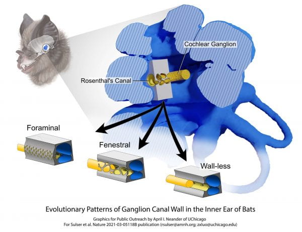
Illustration of anatomy and evolution of bat inner ears: variation of the cochlear ganglion, and its bony tube, inside the inner ear cochlea. Illustration by April I. Neander/UChicago.

First Author Ben Sulser (U.of.C BS 2016) in histological work on bat ear neuroanatomy. Photo courtesy of Ben Sulser.

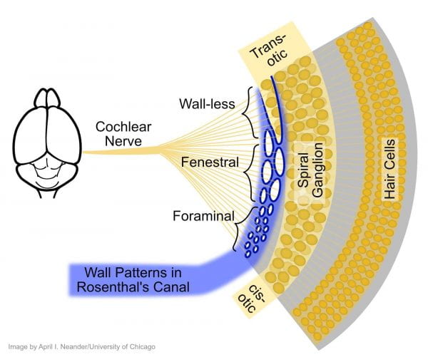
Schematic diagram of the inner ear auditory nerves, and the bony structure (blue) surrounding the spiral ganglion, and the auditory nerve fibers connecting the ganglion to the brain. The nerve fibers and the ganglion have three states of the canal wall in the inner ears of bats. The most diverse yangochiropteran bats are distinctive in having the most derived wall-less pattern. Illustration by April I. Neander/UChicago.

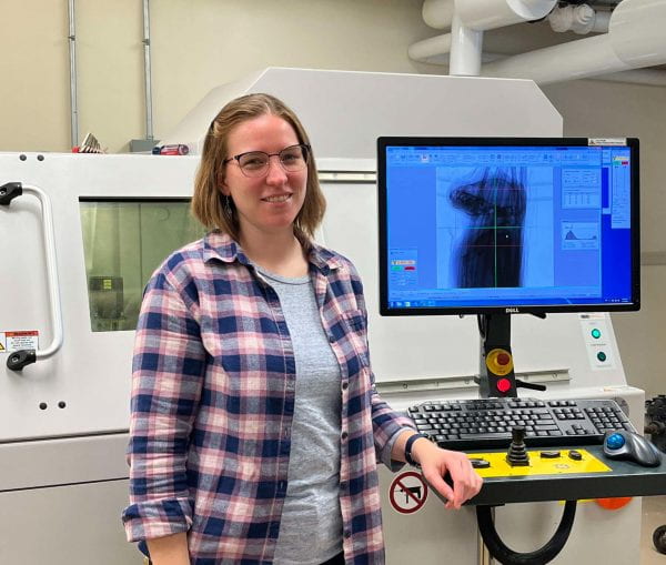
Co-author April I. Neander microCT scanning bat specimen. Photo courtesy of Dr. Zhe-Xi Luo.

Get to know the Authors

Benjamin Sulser, Ph.D. Candidate Richard Gilder Graduate School, American Museum of Natural History, New York, NY, USA

Dr. Bruce Patterson, Negaunee Integrative Research Center, Field Museum of Natural History, Chicago, IL, USA

Dr. Daniel J. Urban, Institute for Genomic Biology, University of Illinois at Urbana-Champaign, Urbana, IL, USA

April I. Neander, Department of Organismal Biology and Anatomy, The University Chicago, Chicago, IL, USA

Dr. Zhe-Xi Luo, Department of Organismal Biology and Anatomy, The University Chicago, Chicago, IL, USA

Scroll to Top