
Read our New paper Evolution of inner ear neuroanatomy of bats and implications for echolocation Here!
Citation:
Sulser, R.B., Patterson, B.D., Urban, D.J. et al. Evolution of inner ear neuroanatomy of bats and implications for echolocation. Nature (2022). https://doi.org/10.1038/s41586-021-04335-z
Research Abstract
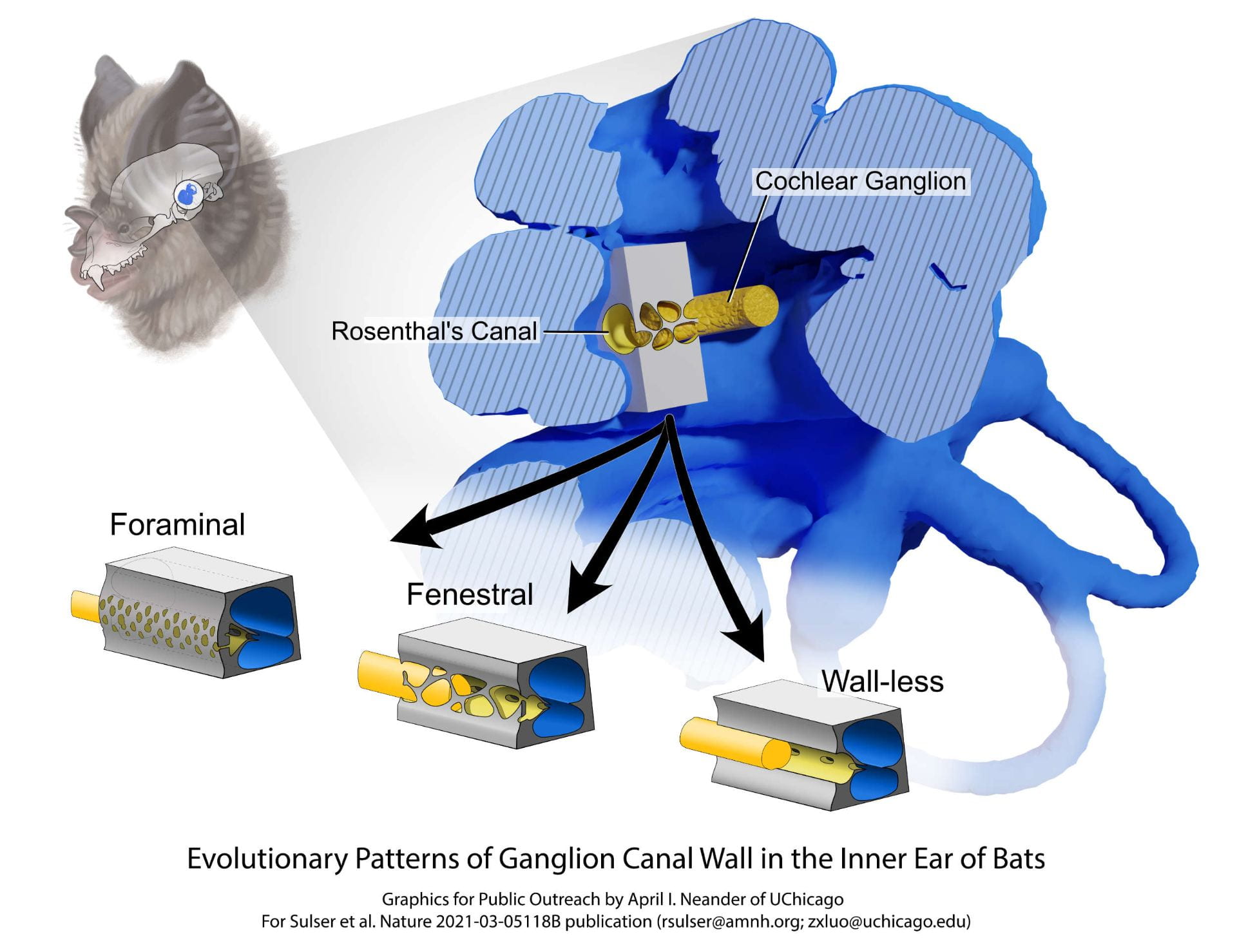
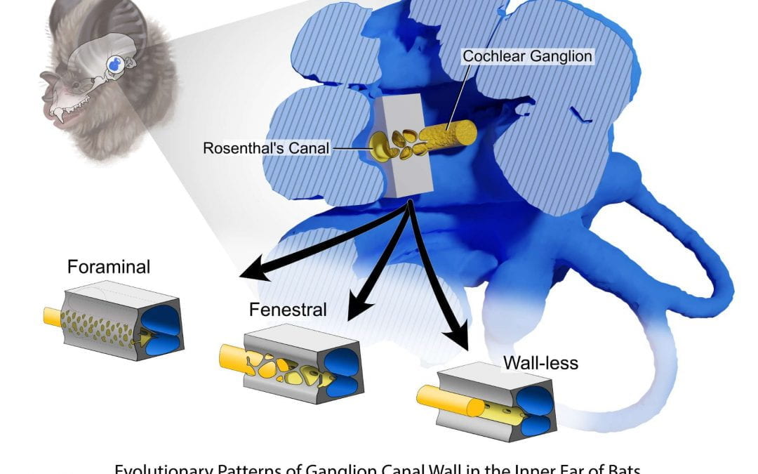
Illustration of anatomy and evolution of bat inner ears: variation of the cochlear ganglion, and its bony tube, inside the inner ear cochlea. Illustration by April I. Neander/UChicago.

First Author Ben Sulser (U.of.C BS 2016) in histological work on bat ear neuroanatomy. Photo courtesy of Ben Sulser.
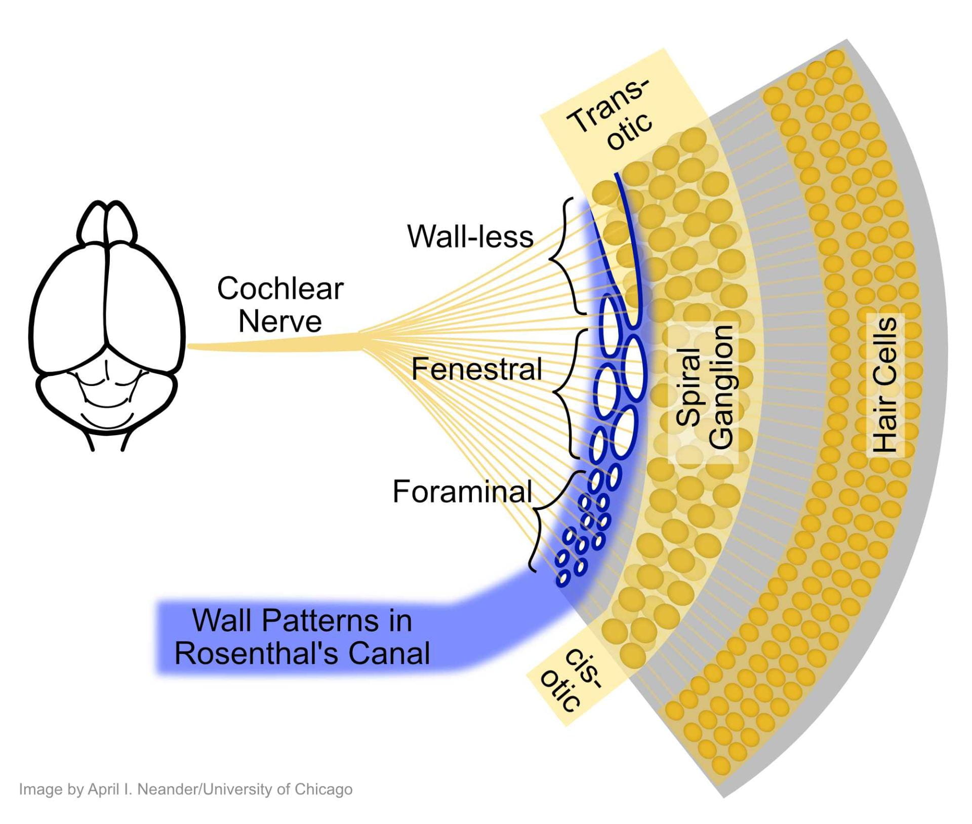
Schematic diagram of the inner ear auditory nerves, and the bony structure (blue) surrounding the spiral ganglion, and the auditory nerve fibers connecting the ganglion to the brain. The nerve fibers and the ganglion have three states of the canal wall in the inner ears of bats. The most diverse yangochiropteran bats are distinctive in having the most derived wall-less pattern. Illustration by April I. Neander/UChicago.

Co-author April I. Neander microCT scanning bat specimen. Photo courtesy of Dr. Zhe-Xi Luo.
Get to know the Authors
Benjamin Sulser, Ph.D. Candidate Richard Gilder Graduate School, American Museum of Natural History, New York, NY, USA
Dr. Bruce Patterson, Negaunee Integrative Research Center, Field Museum of Natural History, Chicago, IL, USA
Dr. Daniel J. Urban, Institute for Genomic Biology, University of Illinois at Urbana-Champaign, Urbana, IL, USA
April I. Neander, Department of Organismal Biology and Anatomy, The University Chicago, Chicago, IL, USA
Dr. Zhe-Xi Luo, Department of Organismal Biology and Anatomy, The University Chicago, Chicago, IL, USA